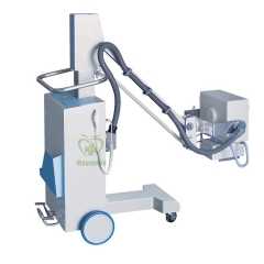
- Description
Specifications
The Unit consists of the following fundamental parts:
♥ tube assembly
♥ generator
♥ collimator
♥ detector
♥ Workstation
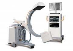
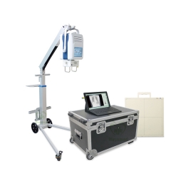
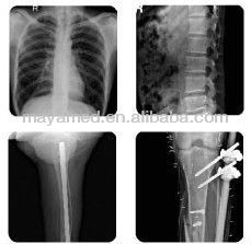
The device works at constant potential high frequency. It connects with standard electrical outlet operation with single-phase at 115/230±10% VAC. During working, adjust the KV value according to different patient; choose the right mA data, X-ray source component transmit corresponding x-ray, which traverses through the patients’ body, producing different attenuation. This attenuated signal is detected by digital detector, processed by workstation, transmitted through Dicom 3.0 interface, output the image of diagnosed part on the screen. The image can be transmitted, seek out, printed and so on. It can provide an aid to diagnosis when used by a qualified physician. The system applies to the hospitals which has a high requirement of digital X-ray examination. It is used in operating room, emergency room and bedside for X-ray radiographic examination. The system can take general X-ray examination of all parts of the body such as breast, head, abdomen, spine and limbs. The system processes, displays, and stores the collected images. The device output can provide an aid to diagnosis when used by a qualified physician.
System Configuration
| Part Name | Quantity |
| High Voltage Generator & X-ray tube | 1 |
| Frame | 1 |
| FP detector | 1 |
| Collimator | 1 |
| Acquisition system | 1 |
| Optional parts | |
| Remote Controlled Handswitch | 1 |
| Vertical bucky stand | 1 |
| Grid | 1 |
System Specification
| Frame | |
| Frame form | Folding |
| Length | Min=1400mm;Max=1834mm |
| Width | 730mm |
| Height | Min=1430mm;Max=2000mm |
| Tube vertical travel range | 525~1992mm |
| The rotating angle | ±90° |
| The maximum distance from center to focus | 1153mm |
| The maximum height of the obstacles | 25mm |
| Control mode | manual |
| X-ray tube | |
| Tube form | combined |
| Power | 30KW |
| Focus | 0.6/1.3mm |
| Anode heat storage capacity | 107KJ |
| Housing heat storage capacity | 375KJ |
| Maximum anode heat dissipation rate | 300W |
| Maximum housing heat dissipation rate | 40W |
| Anode rotation speed | 3000rmp |
| Target Angle | 16° |
| Generator | |
| Rated power | 30KW |
| Inverter frequency | 100kHz |
| kV range | 40~125kV step=1kV |
| Maximum mA | 425mA |
| mAs range | 0.5-200mAs |
| Focal Spots shift | depend on radiography |
| conditions | shifted automatically |
| Time of Radiography | 1ms~1.3s |
| Numbers of APR | 40 (20 for small focus ,20 for big focus) |
| Exposure parameters adjustment method | KV,MAS control |
| Collimator | |
| Adjustment mode | manual |
| Average illuminance | ≥100Lux(SID=1m) |
| Lamp timer | 30S |
| Voltage and power | 12VAC, 9A |
| Inherent filtration | ≥2.0mmAL |
| Light field shape | rectangle |
| Tape measure | With a tape measure SID function |
| Detector | |
| Pixel area | 14X17 inch |
| Conversion screen | Amorphous silicon,DRZ |
| Fill Factor | 100% |
| Image Preview time | 8S |
| A/D output | 14-bits |
| Pixels matrix | 3072 x 2560 |
| Pixels Pitch | 139 um |
| Spatial resolution | 3.6LP/mm |
| Power supply | 100-240VAC,47-63Hz |
| Weight(Including housing ) | about 5Kg |
| Cable length | 8m |
| Imaging working station | |
| CPU | 2.0GHz |
| Memory | DDR2 2G |
| Hard disk | 320G |
| Operating system | WINDOWS XP |
| Peripheral | USB,Mouse,Keyboard ect |
| Screen | 15 inch touch LCD |
| Packing Dimensions(mm) and weight(kg) | |
| Dimension | 1698*858*1662 |
| Weight | 355 |
 USD
USD EUR
EUR GBP
GBP CAD
CAD AUD
AUD HKD
HKD JPY
JPY BRL
BRL KRW
KRW CNY
CNY SAR
SAR SGD
SGD NZD
NZD INR
INR AED
AED MOP
MOP
